BCA

Medical Reports Are Difficult to Read & Grasp

BCA Provides the Clarity You Need for Confident Decision-Making

Understanding Your Reports

Reports Overview

The reports combined give you a picture of the status of your breast cancer and suggest a path for effective treatments. Being willing to pivot in treatment, maybe several times, in order to identify the most effective strategy for you is crucial. 

It is vital that you have these tests run and know your results in order to make good treatment decisions for yourself.

We do not provide medical advice, but we are here to help you understand your reports better through our Patient Advocacy Services service (make this a button link to my calendar)

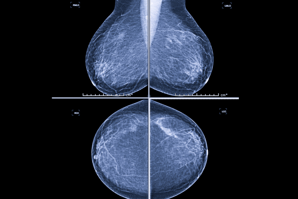
What is a Bi-Rads Score?

Your Mammogram and MRI reports will have a Bi-Rads score on them along with the Radiologist’s findings explaining that score – all in medical lingo. This score is a way to categorize the findings of the scan by the Radiologist. The score is based on the Radiologists’ interpretation of their findings of your likelihood of breast cancer from that scan. . Your ordering physician will go over your results with you and refer you to a breast surgeon if the Radiologist recommends it.


BI-RADS categories 

  • BI-RADS 0: Incomplete, additional imaging is needed Usually attached to Bi-rads 5
  • BI-RADS 1: Negative, no significant abnormalities. Only found for women with no history of  breast surgery
  • BI-RADS 2: Benign, noncancerous growth, such as a cyst or fibroadenoma or women with a history of breast surgery with no suspicious findings because there is scar tissue present. This is the lowest rating for a woman with a history of any kind of breast surgery. Any breast surgery will have scaring and therefore some type of benign findings of abnormalities that are not concerning.
  • BI-RADS 3: Probably benign, but a follow-up study is recommended
    Also for women with a history of breast surgery with no suspicious findings – because there is scar tissue that looks different than normal breast tissue
  • BI-RADS 4: Suspicious, may be cancerous, and requires a biopsy  2%-95% cancer – a very big unhelpful category. That may require a repeat MRI or biopsies depending on the Specialist Doctor’s opinion. Most Breast Oncologist Surgeons and Medical Breast Oncologists read their own Breast MRI’s
  • BI-RADS 5: Highly suggestive of malignancy, requires a biopsy. 
    Most likely breast cancer. This was Roxann’s original MRI finding on her right breast.
  • BI-RADS 6: Known biopsy-proven malignancy, requires treatment.
    This was Roxann’s finding on her left breast after biopsy

These reports can be frustrating to read, especially at Bi-Rads 4 because to a certain extent these are only educated guesses based on an opinion, although educated, and is only a suggestive direction for your doctor. And, if you have been diagnosed with cancer prior and this is a re-staging or treatment surveillance MRI or you are waiting to know if you are diagnosed with breast cancer the waiting is an off the chart anxiety attack for days to a week while you are waiting for the results of your pathology. We recommend you download this Bi-Rads Score Sheet for easy future reference.

BCA Bi-Rads Score PDF →

Pathology Reports

These reports contain a wealth of information about the breast cancer a woman has – and are very complicated to read when you don’t understand the terms and what they mean for you.

We suggest you get yours out, if you have it, make a copy of it –and then be ready to mark it up with a pen so you will remember later what you learned here now.  

What Information does My Pathology Report Contain & Why is it Important?

It first identifies what Type of Breast Cancer you are diagnosed with 

Hormone Receptor Status 

The hormone receptor status of a tumor is crucial. It controls treatment & surveillance:

Treatment Planning: Knowing whether a tumor is hormone receptor-positive (HR+) or hormone receptor-negative (HR-) helps doctors determine the most effective treatment options. HR+ tumors can be treated with hormone therapy, which targets the cancer cells’ dependence on hormones like estrogen and progesterone.

Prognosis: HR+ tumors tend to grow more slowly and respond better to hormone therapy, often resulting in a better prognosis compared to HR- tumors.

Predicting Recurrence: HR+ tumors are more likely to recur, so hormone therapy can be used as an adjuvant treatment to reduce the risk of recurrence after the initial treatment.

Grade of your Tumor

This tells you how much cancer cells look like normal cells under a microscope. It helps predict how quickly the cancer might grow and spread. Grades range from 1 to 3, with Grade 1 looking the most like normal cells and Grade 3 looking the least.

Treatment Planning: The grade helps doctors determine the most appropriate treatment options. Higher-grade tumors (more abnormal cells) may require more aggressive treatment compared to lower-grade tumors (more normal cells).

Prognosis: The tumor grade can give an estimate of the patient’s prognosis. Lower-grade tumors tend to grow more slowly and have a better prognosis, while higher-grade tumors are more likely to grow rapidly and spread, leading to a poorer prognosis.

Behavior Prediction: The grade helps predict how the cancer will behave. Lower-grade tumors are less aggressive and resemble normal cells, whereas higher-grade tumors are more abnormal and tend to be more aggressive

Ki-67 Score

Ki-67 Score: This measures how many cancer cells are dividing. It’s expressed as a percentage, indicating the proportion of cells in the process of dividing1. A higher Ki-67 Score suggests a more aggressive cancer that’s growing quickly.

Assessing Tumor Aggressiveness: The Ki-67 score measures the percentage of cancer cells that are actively dividing. A higher Ki-67 score indicates that the cancer cells are proliferating rapidly, suggesting a more aggressive tumor.

Treatment Planning: The Ki-67 score helps oncologists determine the most appropriate treatment options. For example, tumors with high Ki-67 scores may require more aggressive treatments, such as chemotherapy, while those with lower scores might be treated with hormone therapy.

Prognosis: The Ki-67 score can provide insights into the patient’s prognosis. Higher Ki-67 scores are often associated with a higher risk of recurrence and poorer outcomes, while lower scores suggest a better prognosis..

Monitoring Treatment Response: Changes in the Ki-67 score during treatment can indicate how well the cancer is responding to therapy. A decrease in the Ki-67 score suggests that the treatment is effective in reducing tumor activity.


Tumor Markers

Tumor markers test for specific substances produced by cancer cells or by the body in response to cancer. These substances can be proteins, hormones, antigens, or genetic material. Tumor markers are used by your doctors to:

Diagnose certain types of cancer.

Plan and surveil treatment and check for recurrence

Predict the aggressiveness of the cancer.


My Pathology Report has a Finding of  “Residual Disease” – what exactly does that mean?

When a pathology report mentions “residual disease,” it means that some cancer cells remain in the body after treatment. This can occur after surgery, chemotherapy, radiation, or other treatments. Here are the key points:

  • Residual Disease: Indicates that not all cancer cells were eliminated by the treatment. These remaining cells can potentially grow and cause a recurrence of the cancer.
  • Minimal Residual Disease (MRD): Refers to a very small number of cancer cells that remain, often detected through highly sensitive tests. MRD is commonly used in blood cancers like leukemia.
  • Residual Cancer Burden (RCB): A specific measure used in breast cancer to quantify the amount of cancer remaining after neoadjuvant therapy (treatment given before surgery). The RCB index combines several pathological features to classify the cancer’s response to treatment.

Understanding the presence and extent of residual disease helps you determine your next steps in your treatment and monitor the risk of recurrence. Your doctors of course will have their recommendations, however, the final treatment decisions are always yours to make.

As with most of the other tests described here this test is  not used alone, but in combination with other tests to provide a more complete picture of cancer status because not all breast cancer patients present tumor markers – 20-30% do not, thus they are not reliable as stand alone tests for breast cancer.

Calculation of RCB

The RCB score is calculated using several pathologic features:

  1. Size of the Tumor Bed: The largest two dimensions of the tumor bed in the breast are measured. The tumor bed is the area where the tumor was located and may contain a mix of normal tissue, cancer cells, and scar tissue from the therapy.
  2. Cancer Cellularity: This estimates the percentage of the tumor bed that still contains cancer cells, including both invasive cancer (cancer that has spread into surrounding tissue) and in situ cancer (cancer cells that have not spread).
  3. Percentage of In Situ Disease: Within the tumor bed, the percentage of cancer that is in situ is estimated. In situ cancer cells are confined to the milk ducts or lobules and have not spread into the surrounding tissue.
  4. Lymph Node Involvement: The number of lymph nodes containing cancer cells (positive lymph nodes) is counted, and the size of the largest cluster of cancer cells in the lymph nodes is measured.

These features are combined using a standardized formula to calculate the RCB score.

RCB Scoring and Classification

Based on the RCB score, patients are divided into four categories:

  1. RCB-0 (Pathologic Complete Response): No residual invasive cancer is detected in the breast or lymph nodes. This indicates an excellent response to treatment and is associated with the best prognosis.
  2. RCB-I (Minimal Residual Disease): Very little residual cancer is present. This category indicates a good response to treatment with a favorable prognosis.
  3. RCB-II (Moderate Residual Disease): A moderate amount of cancer remains. This category indicates a partial response to treatment and a moderate prognosis.

RCB-III (Extensive Residual Disease): A large amount of cancer remains in the breast or lymph nodes. This category indicates a poor response to treatment and is associated with a higher risk of recurrence and a less favorable prognosis

Genetic and Molecular Tumor Tests

Onco DX Analyzes the expression of 21 genes to predict the risk of cancer recurrence and the likely benefit of chemotherapy. This is a genomic test used to help guide treatment decisions for certain types of breast cancer.The test is typically used for early-stage, estrogen receptor-positive (ER+), HER2-negative breast cancers that are lymph node-negative or have a small number of positive lymph nodes. This test “scores” the following:

Chance of Cancer Recurrence: The likelihood that the cancer will come back after initial treatment.

Benefit of Chemotherapy: Whether adding chemotherapy or hormone therapy will significantly reduce the risk of recurrence.

Tumor Aggressiveness: The overall aggressiveness of the cancer based on gene expression patterns.

The results provide a score between 0 and 100, which helps doctors tailor treatment plans based on the patient’s individual risk. The lower the score the better the prognosis. The test results, combined with other factors like tumor size, grade, lymph node status, and patient age, help gauge prognosis and treatment plans.

  • MammaPrint: The MammaPrint test is a genomic test that analyzes the activity of certain genes in early-stage breast cancer.
  • Prosigna: a genomic test is intended for post-menopausal women with early-stage HR+/HER2- breast cancer to assess their recurrence risk over 10 years.

Pathology Tests

These tests are another piece of the breast cancer staging puzzle and measure:

  • Histopathology: Examines the tissue sample under a microscope to determine if the tissue sample is benign or malignant. If it is malignant this test determines what type of breast cancer the tissue is.
  • Immunohistochemistry: Tests for specific proteins on the surface of cancer cells to guide treatment options HER2- or +.
  • Hormone Receptor Testing: Checks for estrogen and progesterone receptors to determine if hormone therapy might be effective. This is rated as “ER+” with a % next to it or “ER 0”  and – PR+ with a % next to it or “PR 0”.

This information helps you understand which treatments your breast cancer is likely to respond to and identifies those that won’t be effective for your specific tumor. It guides you and your doctors’ treatment decisions throughout your care and monitoring process.

Breast Cancer Tumor Markers

20-25% of breast cancer patients show no tumor markers at all and some of these tests can be elevated for reasons other than breast cancer.  For this reason these these tests alone cannot be used as a definitive guide one way or the other, the results must be taken together as a whole with other breast cancer surveillance tests and evaluated by your medical team for an interpretation.

  1. CA 15-3 and CA 27.29
    • Function: These markers are proteins produced by breast cancer cells.
    • Use: Elevated levels can indicate the presence of breast cancer or monitor treatment response.
  2. Carcinoembryonic Antigen (CEA)
    • Function: A protein found in some types of cancer cells.
    • Use: Commonly associated with colorectal cancer, but can also be elevated in breast cancer, and can be elevated by non-cancer reasons.
  3. CA 125
    • Function: Typically associated with ovarian cancer.
    • Use: Can also be elevated in breast cancer cases, and can be elevated by non-cancer reasons.
  4. Circulating Tumor Cells (CTCs)
    • Function: Cancer cells that have broken away from the primary tumor and entered the bloodstream.
    • Use: High levels of CTCs can indicate cancer progression and monitor treatment effectiveness.
There will be additional blood tests your doctor(s) will order as part of your big picture.

Other Helpful Blood Tests

There are many other blood tests that give important information to you and your doctors about the status of your cancer and the effectiveness of your treatment:

Inflammation marker

A CRP (C-reactive protein) test measures the level of CRP in the blood, which is a marker of inflammation. Elevated CRP levels can indicate the presence of inflammation, which is often associated with breast cancer progression and poorer prognosis. Here are some key points about the CRP test in cancer patients:

  1. Indicator of Inflammation: CRP levels rise in response to inflammation, which can be caused by cancer or other conditions.
  2. Prognostic Value: Higher CRP levels in cancer patients are often associated with a worse prognosis and more advanced disease.
  3. Site-Specific Associations Elevated CRP levels have been linked to increased risks of certain cancers, including lung, breast, colorectal, and ovarian cancers.
  4. Monitoring Treatment: CRP tests can be used to monitor the effectiveness of cancer treatments, as decreasing CRP levels may indicate a positive response to therapy.

Liver Enzymes

Liver enzyme tests in breast cancer patients are used to monitor liver function and detect potential liver metastasis or damage caused by the cancer or its treatment

Kidney function

Kidney function tests in breast cancer patients are important to monitor the health of the kidneys, especially since certain cancer treatments can affect kidney function

Glucose

Measuring glucose levels in breast cancer patients is important because diabetes and high blood sugar can impact cancer treatment and outcomes.

Estrogen and Progesterone Tests

Monitoring estrogen and progesterone levels & receptors in breast cancer patients is crucial, especially for those with hormone receptor-positive (ER+/PR+) cancers. Here’s why:

  1. Hormone Receptor Status: Breast cancer cells can have receptors for estrogen (ER-positive) and/or progesterone (PR-positive). When these hormones bind to the receptors, they can stimulate cancer growth.
  2. Treatment Decisions: Knowing the hormone receptor status helps doctors decide on the best treatment options. Hormone therapies, such as tamoxifen or aromatase inhibitors, can be used to block the effects of estrogen or lower its levels in the body.
  3. Monitoring Treatment Efficacy: Regularly measuring estrogen and progesterone levels helps assess how well the hormone therapy is working. If hormone levels remain high, it may indicate that the treatment isn’t effective, and adjustments may be needed.
  4. Disease Progression: Changes in hormone receptors can also provide insights into the progression of the disease and help guide further treatment decisions

Chemotherapy Report

After each chemotherapy session, a medical report is typically created and added to your medical records. This report helps your healthcare team monitor your progress, manage side effects, and make informed decisions about your ongoing treatment. This report includes:
  1. Date and Time: When the chemotherapy session took place.
  2. Chemotherapy Drugs Administered: Names and dosages of the drugs given.
  3. Dosage and Administration Details: How the drugs were administered (e.g., intravenously, orally).
  4. Vital Signs: Blood pressure, heart rate, temperature, and other relevant measurements taken before, during, and after the session.
  5. Side Effects: Any immediate side effects experienced during or after the session.
  6. Nurse or Doctor’s Notes: Observations and comments from the healthcare professionals involved.
  7. Plan for Next Session: Recommendations for future treatments, including any changes in dosage or medication.

Radiation Treatment Report

After each radiation session, a medical report is typically created and added to your medical records. This report helps your doctors monitor your progress, manage side effects, and make informed decisions about your ongoing treatment. This report includes:
  • Date and Time: When the radiation session took place.
  • Details of the Session: Information about the type and dose of radiation administered.
  • Positioning and Setup: How you were positioned and any equipment used.
  • Vital Signs: Blood pressure, heart rate, temperature, and other relevant measurements taken before, during, and after the session.
  • Side Effects: Any immediate side effects experienced during or after the session.
  • Nurse or Doctor’s Notes: Observations and comments from the healthcare professionals involved.
  • Plan for Next Session: Recommendations for future treatments, including any changes in dosage or technique.

Operative Reports

Operative reports are detailed documents that capture what happened during a surgical procedure.These reports are crucial for documenting the surgery, supporting medical necessity, and ensuring proper reimbursement from insurance for the facility and medical team. They include:
  1. Facility information: patient details, date of service, and surgery information (surgeon, type of anesthesia, etc.).
  2. History/Indications for Surgery: Explanation of why the surgery was needed and relevant medical history.
  3. Body: Detailed description of the procedure, including steps taken and any complications,
  4. Findings and Follow-Up: Results of the surgery and any follow-up care instructions.

Emotional Support

Good Emotional Support is critical to your healing. Reading your reports is empowering and challenging at the same time. Knowledge is power, that’s true – but it’s stil stressful to read your own cancer reports. The ups & downs from one test to the next takes its emotional toll on you, especially during treatment. Even if the test results are good, it is still stressful getting to that place of peace of mind. Reach out to your support network to help you emotionally often. Then use Duality to fill in the gaps!

References
  • Susan G. Komen, Breaks down the contents of a breast cancer pathology report in detail
  • MyPathologyReports.ca, discusses in detail in the patient sectionBreast
  • Cancer Now, comprehensive booklet on pathology results, helping patients interpret their diagnosis
  • RadiologyInfo.org, how to read breast imaging reports, MRI findings, BI-RADS categories, what different results mean for diagnosis & treatment.
  • Cancer Imaging Journal, Reviews how radiologists report breast MRI studies, staging, screening & the role of MRI in breast cancer management
  • Patient Power, Explains key components to help apaitens understand their chemotherapy results.
  • Darwyn Health, Offers an in-depth explanation of tumor markers & explanations of resultsLANDAUER – Explains how to interpret radiation dose information, including exposure values, unexpected patterns, and discrepancies in reports.
  • LANDAUER – Explains how to interpret radiation dose information, including exposure values, unexpected patterns, and discrepancies in reports.

Subscribe to Our Newsletter to Get the Most Current Breast Cancer Information